
Page 39 - Inaugural Lecture Prof Dr Ahmad Sobri Muda
P. 39
Ahmad Sobri Muda
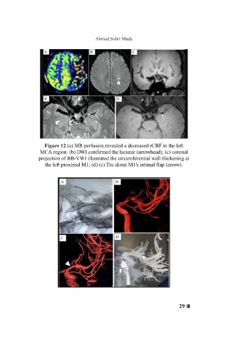
Figure 12 (a) MR perfusion revealed a decreased rCBF in the left
MCA region. (b) DWI confirmed the lacunar (arrowhead); (c) coronal
projection of BB-VWI illustrated the circumferential wall thickening at
the left proximal M1; (d) (e) The distal M1’s intimal flap (arrow).
29 ❘❘❚